What Is Coenurosis?
Coenurosis is a rare parasitic infection affecting the central nervous system. It is caused by a tapeworm called Taenia multiceps which lives benignly in the definitive canine host, such as dogs, foxes, jackals, and coyotes. Coenurosis is a fatal disease affecting sheep and goats. The adult tapeworm grows in the intestines of dogs. In sheep and goats, the larva develop cysts in the brain.
On infecting the central nervous system (CNS), the organism produces thin, translucent, whitish, and large uni or multilocular cysts, measuring up to 10 cm in diameter. The cysts contain clear, watery fluid with hundreds of white nodules on their inner surface, measuring a few millimeters in diameter (Benifla et al., 2007).
The nodules correspond to multiple protoscolices that develop from invaginations of the germinative layer of the cyst. A rostellum with rows of hooklets can be identified in each protoscolex. The cyst is surrounded by chronic inflammatory reaction (lymphocytes, plasma cells, and foreign-body giant cells), fibrosis, and gliosis. The leptomeninges, brain parenchyma and ventricles are the preferred sites of cysts.
Sheep are the main intermediate hosts. But rarely, cattle, pigs, deer, horses, and humans serve as intermediate hosts. This tapeworm causes significant problems in the intermediate host when the larval stage of the tapeworm migrates, matures, and forms a fluid-filled cyst in the brain and spinal cord.
The prevalence of coenurosis is challenging to assess because farmers and veterinarians often diagnose the disease and send the animal for slaughter without any confirmation. Many infected lambs are also sold with fat before any clinical signs are developed.
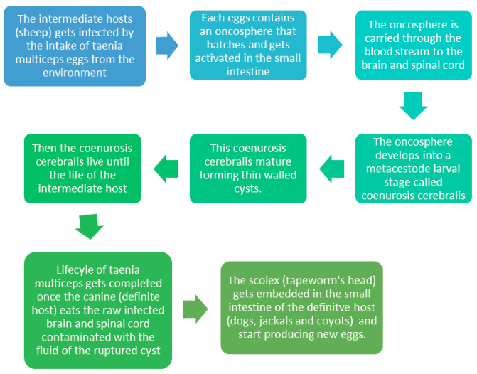
What Is the Life-Cycle of Taenia Multiceps?
The definitive hosts for Taenia multiceps and Taenia serialis are members of the family Canidae. Many canids can serve as definitive hosts for T. multiceps, but only dogs and foxes can serve as hosts for T. serialis. Eggs and gravid proglottids are shed in feces into the environment, where an intermediate host ingests them. Many animals may serve as intermediate hosts, including rodents, rabbits, horses, cattle, sheep, and goats.
Eggs hatch in the intestine, and oncospheres are released that circulate in the blood until they lodge in suitable organs (including skeletal muscle, eyes, brain, and subcutaneous tissue). After about three months, oncospheres develop into coenuri. The definitive host becomes infected by ingesting the tissue of an infected intermediate host containing a coenurus. The adult cestodes reside in the small intestine of the definitive host.
Humans become infected after accidentally ingesting eggs on fomites or in food and water contaminated with dog feces. Eggs hatch in the intestine, and oncospheres are released that circulate in blood until they lodge in suitable organs and develop into coenuri after about three months. Coenuri of T. multiceps are usually found in the eyes and brain; those of T. serialis are usually found in subcutaneous tissue.

What Are the Clinical Signs of Coenurosis?
Signs of coenurosis begin to occur when the central nervous system of the sheep is infected with the metacestode larval stage of the tapeworm. Coenurosis can occur in acute and chronic forms.
The symptoms of acute coenurosis are shown in the sheep after ten days of ingesting many tapeworm eggs. The common signs shown are inflammation, allergic reactions, and transient pyrexia. Mild neurological symptoms with fatigue and head aversion are also shown. Young lambs aged six weeks to eight weeks are more likely to show acute coenurosis.
In some cases, the signs are more severe, and the affected animal develops encephalitis, convulsions and dies within four to five days. This acute coenurosis also serves as an important differential diagnosis for cerebrocortical necrosis.
Chronic coenurosis occurs in sheep of sixteen to eighteen months of age. The early sign shown in the affected animal is a behavioral change. The time taken for the larva to hatch, migrate and grow larger to present a neurological dysfunction varies from two months to six months.
The affected animal may stand away from the flock and react slowly to external stimuli. On the growth of the cyst, the clinical signs shown in the affected animal are depression, unilateral blindness, paralysis, altered head position, and incoordination. Unless treated surgically, the affected animal will die.
How Does Coenurosis Affect Humans?
Humans are occasionally and accidentally infected with the metacestode (juvenile stage) of Taenia multiceps, T. brauni, T. serialis or T. glomerata, the adult forms of which normally infect dogs. Intermediate hosts include cattle, sheep and rodents. Metacestodes, known as coenuri, are released from eggs ingested in contaminated water or food.
These penetrate the intestinal wall and migrate to subcutaneous tissues and in the case of T. multiceps, to the central nervous system (CNS) where they can form large cysts, usually 2-5 cm in diameter, with multiple invaginated protoscolices.
CNS disease can present with headache, seizures, hemiparesis, or hydrocephalus, and is frequently fatal. Human cases are rare and largely occur in Africa, although infections have been reported worldwide. Coenurosis symptoms may take several years to develop and depend on the organ infected.
Coenurosis infection affecting the brain causes increased intracranial pressure, seizure, loss of consciousness, and focal neurologic deficits. The coenurus infection in the subcutaneous tissue manifests as a tender nodule. When the eyes get infected, impairment in vision occurs.
How to Diagnose and Treat Coenurosis in Humans?
Diagnostic criteria include epidemiologic, clinical, radiologic and biopsy findings; serology is unavailable. Diagnosis of coenurosis is made after the surgical removal of coenurus is done in symptomatic cases. Treatment usually involves surgical excision if the cyst is accessible, although anthelmintic therapy with albendazole and Praziquantel has been successful in some patients. Praziquantel is not advisable for people with intraocular coenurosis as the dying parasites can cause severe inflammation resulting in vision loss.
How to Treat Coenurosis in Animals?
Currently, the only treatment recommended is the surgical removal of the coenurus cyst from the brain of the affected animal. This treatment seems successful, and the affected animal may show complete recovery with normal neurological function. But surgery is not advisable for all the affected animals because surgery is advised based on the location of the cyst. The veterinarian doctor will decide whether the affected animal will recover or if it is good to destroy the animal to avoid further suffering.
How to Control and Prevent Coenurosis?
Coenurosis is controlled and prevented by preventing dogs from accessing sheep and cattle carcasses and avoiding the feeding of uncooked meats. If this measure does not help, the control and prevention of coenurosis should be made by routine anthelmintic dosing of dogs, preferably every three months.
Public footpaths running through the sheep fields used by people walking with their dogs can also create problems. Farmers could display awareness among people regarding the disease risk. Farmers may also help people understand that walking with their dogs in these fields may get infected with coenurosis. To identify coenurosis and other ailments, the farmer should regularly invite a veterinarian to examine the livestock.
What Are the Steps to Be Followed to Avoid Coenurosis in Farms?
When coenurosis occurs as a general occurrence in the farm, eliminating the disease from the farm should be a part of the overall health plan. Efforts must me made to prevent dogs and other canines from contaminating pasture with tapeworm eggs by stopping them from eating sheep carcasses. Sheep carcasses should be disposed of quickly and correctly. If possible, local people walking with dogs on the land should be encouraged to deworm their dogs, preferably once in three months.
Conclusion:
Like other well known metacestodes, Coenurosis caused by T. multiceps is a significant health concern that can lead to notable economic losses in small ruminant breeding and also constitutes a non-negligible zoonotic risk. Thus, it is appropriate to conduct further research on chemical prophylactic protocols for coenurosis (vaccines and drugs) and alternative strategies for its control, such as preventing infection in definitive hosts.
The valorization of sheep products (and goat products) could also help to promote effective measures against these metacestodes. This coenurosis creates disastrous health issues in animals as well as humans. Preventing sheep from being infected with coenurosis is the utmost step to be followed to save all animals and humans infected with this disease.


