Patella is a small oval-shaped bone in the trochlear groove in front of the knee, which covers and protects the knee joint. It stabilizes the quadriceps muscles and connects the thigh muscles with the tibia or the shin bone. It has a thick layer of cartilage on the inner side, which cushions and helps in the smooth gliding movement of the knee joint. Any damage to the cartilage can result in post-traumatic arthritis, or any defect or damage to the trochlear groove leads to sliding out of the knee cap from the groove (subluxation) or complete dislocation of the patella.
What Is a Kneecap Fracture?
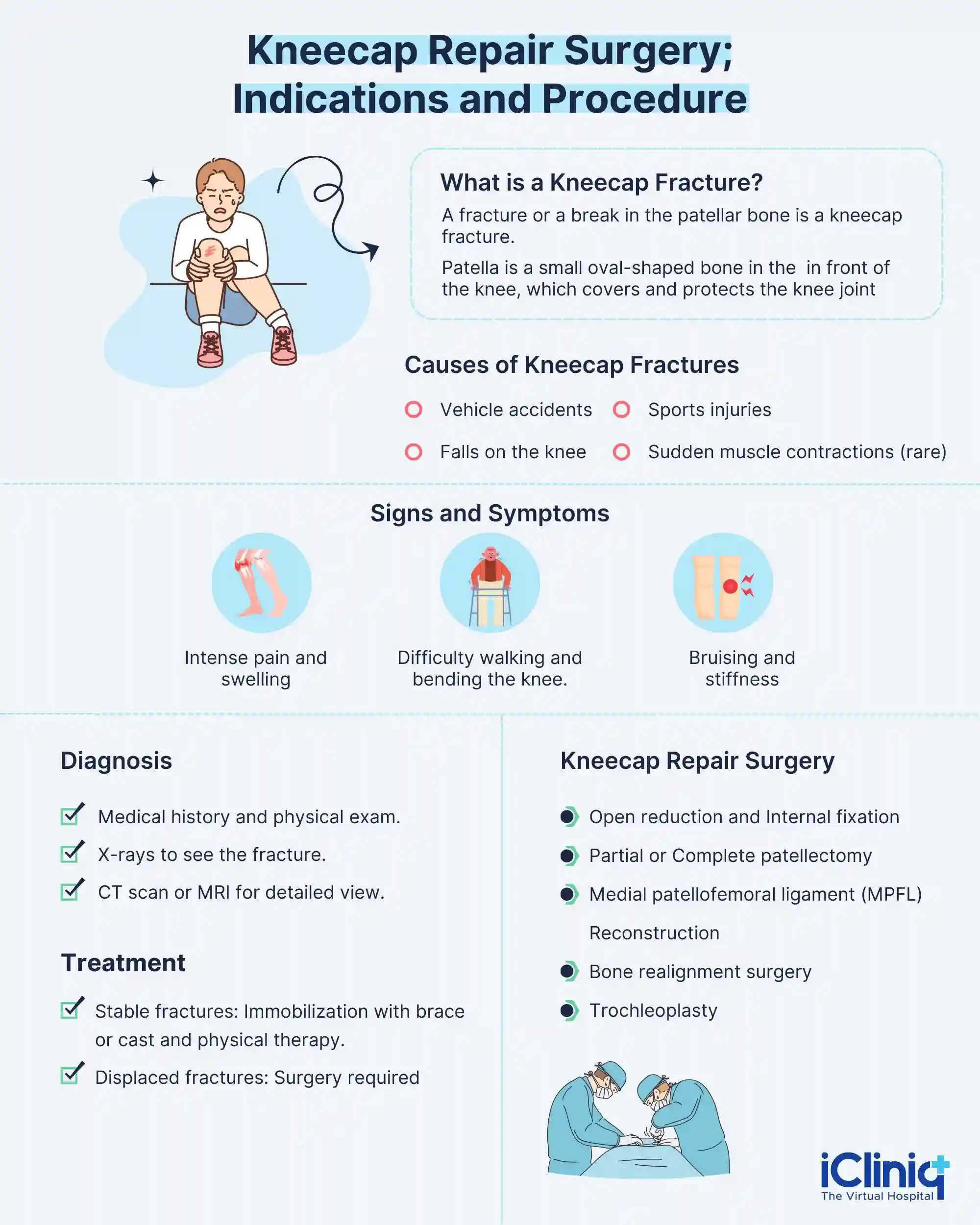
A fracture or a break in the patellar bone is a kneecap fracture. It is a serious injury that may result when the kneecap may be broken into several fragments, or it may just be a crack in the bone, depending on the type of injury and quality of the bone. The quadriceps muscle and the patellar tendon help in the bending and straightening movements of the knee.
What Are the Types of Kneecap Fractures?
Different types of patella or kneecap fractures include
-
Stable Fracture: These are nondisplaced fractures wherein the fracture fragments remain in place or may be separated by a few millimeters. These fractures heal by immobilization in the extension position, with a knee immobilizer, a hinged knee brace, or by cast application.
-
Displaced Fracture: In these types of fractures, the fracture fragments are separated and displaced from their positions. It requires surgical management in order to heal and promote proper functioning of the joint.
-
Transverse Patellar Fracture: In this type of fracture, the patellar bone is broken into two pieces and is usually managed by surgical treatment.
-
Comminuted Patellar Fracture: These types of fractures occur due to severe injuries, breaking the bone into many fragments; some pieces may be too small to reconnect and require surgical removal.
-
Open Patellar Fracture: The skin over the bone is brokenin this type of fracture, and the bone is exposed. It requires immediate management by cleaning the wound and antibiotics administration, followed by surgical treatment to prevent infections.
What Are the Causes of Kneecap Fractures?
Some of the causes of kneecap fractures include:
-
Vehicle accidents and dashboard injuries cause a direct blow to the knee.
-
It can be caused due to hard fall on the knee or sports injuries that occur during football, athletics, etc.
-
In some rare cases, due to sudden muscle contraction in the knee.

What Are the Signs and Symptoms of Kneecap Fractures?
Some of the signs and symptoms of kneecap fractures include:
-
Intense pain immediately following the injury.
-
Pain worsens during knee movement or contraction of thigh muscles.
-
Development of swelling within a few hours of injury.
-
Inability to walk.
-
Inability to straighten or bend the knee.
-
Bruises around the knee joint.
-
Unable to balance the body weight.
-
Stiffness of the knee joint.
What Are the Complications of Kneecap Fractures?
Some of the complications of kneecap fractures include:
-
Severe kneecap fractures can damage the joint cartilage, leading to inflammation of the joints, called post-traumatic arthritis.
-
Chronic pain and permanent weakness of the quadriceps muscles lead to difficulty bending and stretching the knees.
-
Post-surgical complications include infection and delayed wound healing may be seen.
How Is a Kneecap Fracture Diagnosed?
A complete medical history is taken following a complete physical examination of the knee. Skin abrasions, bleeding, lacerations and bruises are observed in fractures due to trauma. In cases of displaced fractures, the fractured edges are felt through the skin. In the presence of collection of blood in the joint (haemarthrosis), it is drained to reduce pain and swelling. Radiological investigations include X-rays to determine the type, severity, and extent of fractures. In some cases, a computed tomography (CT Scan) or magnetic resonance imaging (MRI) may be recommended to evaluate soft tissue injuries.
How Is a Kneecap Fracture Managed?
Stable or undisplaced fractures are managed by immobilization with a hinged knee brace or cast for around four to six weeks, along with medications like non-steroidal anti-inflammatory drugs. It is followed by physical exercises and rehabilitation therapy. Displaced, transverse, and comminuted open fractures have to be managed by surgical treatment. Individuals with recurrent patella instability or failed closed reduction cases may also require surgical management.
What Is Kneecap Repair Surgery?
The surgery that is performed to repair a fractured and displaced kneecap or patella is the kneecap repair surgery. The type of surgical treatment depends on the type of fracture. Surgical treatment methods include:
-
Open Reduction and Internal Fixation (ORIF): ORIF or Open reduction is an open surgery performed to align the fractured fragments into their normal position. Internal fixation is fixing the aligned fragments in position to prevent displacement. It is done with the help of metal screws, plates, or wires to facilitate healing. Immediate treatment with ORIF reduces the pain, swelling, and discomfort and aids in healing, and prevents further complications. The surgery is performed under general anesthesia, with all aseptic precautions. An incision is done at the fracture site, and the area is exposed; very small bony fragments that cannot be fixed are removed. The bones are then aligned and fixed using metal screws or plates. The surgical area is closed with the help of sutures or staples. A sterile dressing is done and immobilized with a cast.
-
Partial or Complete Patellectomy: It is recommended in cases of severely comminuted fractures which cannot be managed by ORIF. It involves partial or complete removal of the patellar bone to relieve the symptoms and restore function. During this procedure, an incision is made on the quadriceps tendon, and the patella is set free and removed; the patellar tendon is intact and partially functions. It helps preserve the quadriceps tendon, patellar tendon below, and surrounding soft tissues. This is followed by rehabilitation to promote healing and restore normal function.
-
Medial Patellofemoral ligament (MPFL) Reconstruction: Repair and reconstruction of the medial patellofemoral ligament is done in cases of surgical management of patellar instability. The patient is asked to lie in a supine position, and a well-padded high-thigh tourniquet is applied to the operative leg. The tendon of the Gracilis muscle is removed by skin incision anteromedially exposing the upper border of the patella. V-shaped tunnels are drilled on the inner aspect, and a second tunnel is drilled where the original ligament is attached to the thigh bone. Around ten millimeters of cortical bone between the two tunnels is left behind to avoid fracture. To reconstruct the MPFL, a single hamstring tendon is used, which is passed between the two tunnels as a loop and secured in position.
-
Bone Realignment Surgery: It is recommended in cases of anatomical abnormalities of the patella, which causes instability. It involves detaching the tendon, along with a small block of bone, and is moved towards the midline. It is then fixed with metal screws, and the position is secured.
-
Trochleoplasty: It is a surgical procedure that is advised in case of abnormal trochlear groove (trochlear dysplasia), which reshapes the groove and allows the smooth movement of the patella, preventing instability and pain. Ligament construction may be done in some cases following trochleoplasty.
Conclusion
Surgery of the kneecap or the patella bone is performed in cases of comminuted or displaced fractures or severe cases of patellar instability or anatomical abnormalities. It is done to reduce the symptoms and restore the function of the kneecap. ORIF is the most common method to treat patellar fractures. Appropriate surgical treatment and rehabilitation by physical exercises improve the range of motion and strengthen the muscles.


