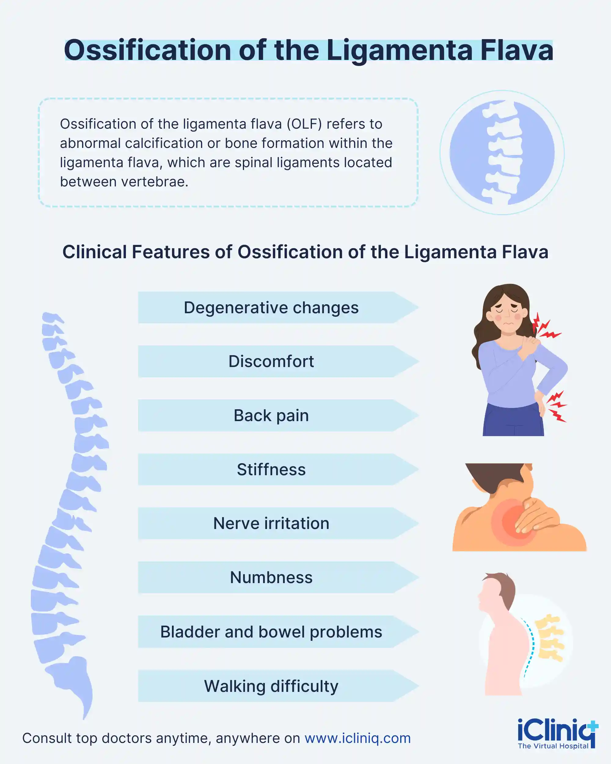
What Is Ossification of the Ligamenta Flava?
Ossification of the ligamentum flavum is a rare condition, and its cause is not fully understood yet. However, it seems to be related to changes in spine mobility, especially tension changes. It is commonly found in the lower thoracic level, causing many neurological symptoms that are determined by the degree of compression on the nerve roots, spinal cord, cauda equina (group of nerves and nerve roots arising from the distal portion of the spinal cord), and conus medullaris (lowermost tapering of the spinal cord). Polgar initially discovered ossification of the ligamentum flavum (OLF) using lateral X-ray films, but serious research into the condition began after the emergence of MRI (magnetic resonance imaging) and CT (computed tomography).
The ligamentum flavum connects two adjacent laminas behind the dura mater, with its side flaps separated at the midline. It extends to the front of the facet joint and is separated from the dura mater by epidural fat. This condition compresses the spinal cord from the back, resulting in walking difficulties and stiffness in the legs that gets worse as the compression increases. There are relatively few cases of OLF in Western countries, leading to ossification of the ligamentum flavum and ossification of the posterior longitudinal ligament (OPLL), being colloquially referred to as "Japanese diseases."
What Is the Etiology of Ossification of the Ligamenta Flava?
The cause of ossification of the vertebral ligaments remains unclear, but it can either be due to systemic or local factors. Systemic factors may involve abnormal calcium metabolism, abnormal carbohydrate metabolism, abnormal hormone secretion, heredity, and ligament degeneration. Local factors comprise mechanical stress at the point where the ligament attaches to the bone, particularly at the capsular portion.
At the lower thoracic level, where the spine has a kyphotic alignment, the ligamentum flavum is subject to strong traction force due to rotational flexion movements of the thoracolumbar spine, as it is located far from the center of the movement. This traction force is thought to have a role in the ossification process. Conversely, in the ligamentum flavum of the lumbar and cervical spines, where the spine has a lordotic alignment, calcification without trabecular structure can occur rather than ossification. It is believed that, at these levels, the traction force does not act strongly. This distinction is important in understanding calcium metabolism in relation to the spinal alignment, vertebral level, and the strength of traction force.
What Are the Clinical Features of Ossification of the Ligamenta Flava?
-
Ossification of the ligamenta flava is a degenerative change commonly seen with aging in the spinal column.
-
Patients with ossification of the ligamenta flava usually experience tight, constricting discomfort, which may result from irritation of the intercostal nerves by the OLF itself.
-
Also, affected individuals may suffer from back pain, back muscle stiffness, and pain during movement due to degenerative changes in other spinal column structures.
-
Systemic symptoms such as pain, numbness, walking difficulty, lower extremity weakness, and bladder and bowel problems, as well, can occur.
-
These symptoms occur as a result of compression of the spinal cord and spinal nerves by the OLF.
-
Ossification of the ligamenta flava usually develops in the lower thoracic region, where the spinal cord transitions to the conus medullaris and the cauda equina.
-
The specific symptoms experienced depend on which nerve tissues are affected, including spastic or flaccid paralysis in the lower extremities, conus medullaris syndrome (lesions around vertebral L2 level), supra-conus medullaris syndrome (lesions around vertebral L2 level affecting the conus medullaris).

How Is Ossification of the Ligamenta Flava Diagnosed?
While the level of nerve tissue compression is often considered the most indicative factor, diagnosis can be challenging in specific cases. However, diagnosis can be challenging in certain cases. Doctors usually advise an MRI scan to take the spine closely. A whole spine scan might also be suggested for identifying issues in the lower back. CT scans will be required for a detailed analysis and to decide the surgical plan.
How to Manage Ossification of the Ligamenta Flava?
-
Rest and physical therapy are not very effective for this condition.
-
Surgery to decrease compression in the spinal cord, known as decompression surgery, is considered the best treatment approach.
-
Prognosis is also better in this approach as compared to other issues in the thoracic region of the spinal cord.
-
Conservative treatments like corset use have shown limited effectiveness.
-
Early surgical intervention is strongly suggested for patients exhibiting a loss of strength in lower limbs, bladder and bowel dysfunction, and spastic gait (walking pattern in which the affected individual drags both legs and scrapes the toes).
-
Also, surgery is crucial in serious compression of the spinal cord due to thickened ligaments and ossification of another ligament in the same area.
-
Symptoms like numbness in the lower extremities and persistent spastic gait show little improvement, which persists even after surgery.
-
So, it is essential to obtain informed consent from patients and their families regarding the potential outcomes of the surgery before the start of the surgery.
-
Very rarely, surgical complications such as CSF (cerebrospinal fluid) leakage, immediate postoperative neurological deterioration, dural tear, epidural hematoma (bleeding between the inside of the skull and dura mater), and wound infection have occurred.
-
As a result, in some cases, the arachnoid membrane is unintentionally ruptured during OLF.
-
Notably, there have been definitive recurrences of OLF at the same intervertebral level where previous decompressions for OLF were performed.
Conclusion
In recent times, surgery has become a safer option due to advances in preoperative imaging technologies, improved surgical skills, and the use of intraoperative navigation systems. However, accurately diagnosing the condition of adhesion between OLF and the dura mater, as well as the presence of ossified dura mater, remains challenging prior to surgery. Clinicians are advised to perform CT imaging where there is a suspicion of OLF based on MRI findings, as it will aid in better preoperative surgical planning.


